
In the field of dermatology, CHDR has developed a unique approach to early-stage drug development by integrating outpatient trials with cutting-edge measurement techniques. In addition to capturing patients' subjective experiences, such as pain, discomfort, and ease of application, we also employ objective measurements to evaluate the effectiveness of new dermatological treatments. By combining a range of proven, reliable techniques commonly used in dermatology research, we have created the innovative DermaToolbox to provide comprehensive insights into treatment outcomes.
An overview of the DermaToolbox tests
-
Laser Speckle Contrast Imaging (LSCI) is a non-invasive technique used to assess cutaneous microcirculation, offering high spatial and temporal resolution. It works by illuminating tissues with laser light, creating a speckle pattern from the interference of reflected light. This pattern is influenced by the movement of red blood cells, with changes in blood flow or cell concentration altering the speckle pattern. LSCI captures these changes, providing a quantitative measure of tissue perfusion. Due to its fast measurement time, LSCI reduces sensitivity to motion artefacts, ensuring reliable and repeatable results, making it an effective tool in pharmacological research and the study of microvascular function.
LSCI applied:
A multimodal, comprehensive characterization of a cutaneous wound model in healthy volunteers. PMID: 37051698
OX40L Inhibition Suppresses KLH-driven Immune Responses in Healthy Volunteers: A Randomized Controlled Trial Demonstrating Proof-of-Pharmacology for KY1005. PMID: 35092305
A randomized controlled trial with a delayed-type hypersensitivity model using keyhole limpet haemocyanin to evaluate adaptive immune responses in man. PMID: 33025648
Extending the IMQ Model: Deep Characterization of the Human TLR7 Response for Early Drug Development. PMID: 39183259
-
At CHDR, we use multispectral imaging with the handheld Antera 3D camera to capture high-resolution images of skin conditions. This innovative technology employs advanced optical methods and mathematical algorithms to create three-dimensional images of the skin. By providing detailed data on the skin's shape and structure, the Antera 3D camera allows us to quantify the efficacy of treatments and track changes in skin condition over time, offering valuable insights for clinical research and dermatological studies.
Multispectral imaging applied:
Oral prednisolone suppresses skin inflammation in a healthy volunteer imiquimod challenge model. PMID: 37545524
Omiganan Enhances Imiquimod-Induced Inflammatory Responses in Skin of Healthy Volunteers. PMID: 32043302
Comprehensive, Multimodal Characterization of an Imiquimod-Induced Human Skin Inflammation Model for Drug Development. PMID: 29768709
Clinical, Cellular, and Molecular Effects of Corticosteroids on the Response to Intradermal Lipopolysaccharide Administration in Healthy Volunteers. PMID: 34935141
-
Transepidermal Water Loss (TEWL) is a key indicator of skin barrier function, reflecting the amount of water that evaporates from the skin's surface. A healthy skin barrier prevents excessive water loss and protects against external irritants. When the barrier is compromised, as seen in conditions like psoriasis, eczema, and atopic dermatitis, TEWL levels increase. At CHDR, we use the Aquaflux and GPSkin Barrier Pro-1, non-invasive devices, to measure TEWL. This tool helps assess skin condition and can aid in the early identification or prediction of skin disorders, making it valuable in both clinical and research settings.
TEWL applied:
A multimodal, comprehensive characterization of a cutaneous wound model in healthy volunteers. PMID: 37051698
A suction blister model to characterize epidermal wound healing and evaluate the efficacy of the topical wound healing agent INM-755 in healthy volunteers. PMID: 39084539
Topical antimicrobial peptide omiganan recovers cutaneous dysbiosis but does not improve clinical symptoms in patients with mild to moderate atopic dermatitis in a phase 2 randomized controlled trial. PMID: 33010325
-
We utilise an automated, standardized method for total body imaging, providing precise and reproducible mapping of the entire body. This technique allows for comprehensive monitoring of dermatological diseases over time, capturing high-quality images to assess skin conditions and track their progression.
-
Target lesion quantification is an objective, computer-based method used to assess the extent and severity of skin lesions, particularly in dermatological conditions like psoriasis. By using whole body photographs, advanced software quantifies the percentage of affected skin, offering a more reliable and reproducible alternative to traditional clinical assessments. This semi-automated approach reduces subjectivity, providing a consistent way to monitor treatment effects and track disease progression over time
-
Optical Coherence Tomography (OCT) is a non-invasive imaging technique used to visualize skin morphology and structure up to 2mm beneath the surface. Using the VivoSight Dx OCT scanner, reflected light from the skin creates detailed images by measuring light refraction, allowing for the differentiation of various skin structures. This method, enhanced with software algorithms, can also visualize cutaneous microcirculation, providing valuable insights into the skin's condition and aiding in skin characterization for research and clinical applications. OCT is a powerful tool in dermatology, offering high-resolution, real-time imaging without the need for biopsies.
-
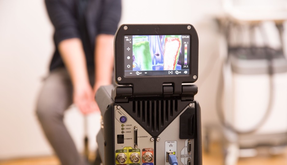
Thermography is a valuable tool for assessing skin temperature, which can indicate the inflammatory status of the skin and serve as a potential biomarker in wound diagnostics. Using the X6540sc thermography camera, infrared technology detects the release of heat from the skin and converts it into an electronic signal to create a thermal image. This allows for the visualization and quantification of temperature changes, often associated with inflammation. Thermography is particularly useful for monitoring wound healing and inflammation, providing precise, non-invasive measurements that support clinical decision-making.
-
3D imaging is a powerful tool for the accurate quantification of skin lesions, offering a more reliable method for evaluating disease severity and assessing therapeutic response. Using the 3D LifeViz Micro™ system, we capture high-quality, standardized images of skin lesions through a dual beam camera. The system, combined with DermaPix® software, allows for the precise analysis of lesion size and skin area roughness. This non-invasive method provides valuable insights into changes in skin morphology over time and is commonly used in clinical studies to monitor the progression or improvement of skin conditions.
-
In addition to advanced imaging techniques, we incorporate clinical scores in our dermatology trials to assess the severity and progression of skin conditions. These scores, performed by trained clinicians, evaluate key factors such as lesion size, erythema, thickness, and scaliness. Clinical scoring methods, including widely recognised indices like the Psoriasis Area and Severity Index (PASI), provide a standardised approach to track treatment efficacy and disease improvement over time, complementing objective measurements and enhancing the overall assessment of therapeutic responses.
The DermaToolbox in action
Watch this video to get an overview of the main DermaToolbox tests: