Executive Summary
Central nervous system drug development faces a recurring problem: when a programme fails, researchers are often left uncertain whether the biological hypothesis was incorrect or whether the research never generated sufficient evidence to test the compound properly.
Plasma exposure and tolerability do not prove that a drug entered the brain, bound its intended target, modulated the relevant pathway, or changed the circuit expected to drive benefit. In an analysis of 44 Phase 2 decisions, 43% of failures could not establish whether the mechanism had been adequately tested - a problem the authors addressed through the integrated "three pillars" of site-of-action exposure, target binding, and pharmacological activity.1
Neuroimaging can close several of those evidence gaps, but only when the modality is matched to the biological question. PET can provide direct or near-direct evidence of molecular distribution, occupancy, or target-related pathology when a suitable radioligand exists. MRI is generally less molecularly specific, but can provide repeatable downstream measures of central pharmacology, physiology, structure, and safety. The value of either modality depends less on the scanner itself than on the decision it is designed to support.
The distinction matters. Strict target engagement is the physical interaction between a compound and its target. A change in BOLD connectivity or perfusion is generally not proof of molecular occupancy. It is evidence of a circuit-level or physiological response that becomes persuasive when it is temporally linked to exposure, moves in the predicted direction, demonstrates dose-response, survives placebo and physiological controls, and converges with a more proximal biomarker.
Specialised CROs have helped build this evidence-based approach through clinical pharmacology studies that combine tightly controlled drug administration with PET, ASL, resting-state fMRI, psychometric and neurophysiological testing, and PK/PD analysis. Examples include measurement of THC-induced striatal dopamine release with 18F-fallypride PET; separation of morphine and alcohol effects on cerebral perfusion with PCASL; mapping of linear and nonlinear dopaminergic effects on resting-state networks; and cross-compound linkage of psychomimetic perception to functional connectivity.3-7
The practical message for sponsors is straightforward: do not add imaging as an exploratory ornament. Start with the decision that must be made, define the biological link the scan is intended to test, design the exposure and controls around that link, and specify in advance what result will advance, redirect, or stop the programme.
Reframe CNS target engagement as a chain of evidence
The phrase “target engagement” is often stretched until it covers any post-dose change in the brain. That imprecision can obscure rather than clarify development risk. Building on the three-pillar framework of site-of-action exposure, target binding, and pharmacological activity, early CNS studies should treat target engagement as one link in a broader evidence chain. The key question is whether the compound reaches the relevant brain compartment, interacts with the intended target, produces the expected pathway or physiological response, and generates a signal that is consistent with the intended functional or clinical effect.
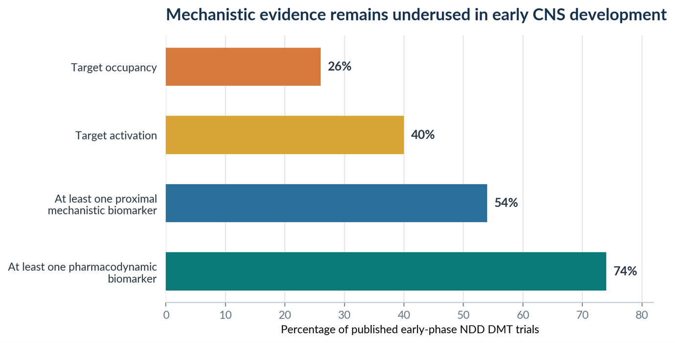 Figure 1. A review of 121 published early-phase trials of disease-modifying therapies (DMT) in neurodegenerative disorders (NDD) found broad biomarker use but substantially lower use of target occupancy and target activation measures. Source: adapted from Vissers et al.2
Why this gap matters
Early trials are often underpowered and too short to establish clinical benefit. That makes mechanistic evidence essential: it can show whether the compound has biological activity in humans before clinical efficacy can reasonably be tested. No single study needs to answer every question. The objective is to establish whether the mechanism has been tested well enough to support a go/no-go decision, dose selection, or further investment. This can prevent later-stage trials from being built on an unresolved biological question.
For a well-characterised receptor with a validated tracer, a PET occupancy study may be decisive. For an intracellular target without a tracer, the package may instead combine CSF exposure, a peripheral or CSF pathway biomarker, and a mechanism-anchored MRI response. For a disease-modifying biologic, pathology PET and fluid biomarkers may establish biological activity while structural MRI provides essential safety monitoring.
This hierarchy also prevents overclaiming. PET competition studies can be close to direct engagement. MRS may provide pathway-proximal chemistry when the targeted mechanism predicts a specific metabolite shift. Resting-state fMRI and ASL usually sit further downstream. They can be highly sensitive to central drug effects, but their interpretation depends on exposure timing, arousal, motion, cardiovascular and respiratory changes, analysis choices, and the specificity of the experimental perturbation.3,4
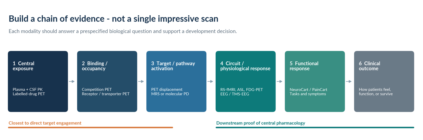 Figure 2. A modality becomes decision-enabling when it is assigned to a defined link in the evidence chain and integrated with study design, exposure control, challenge models, physiological monitoring, and PK/PD modelling.
Target engagement is necessary, not sufficient
A positive occupancy or pathway signal establishes that the mechanism was tested. It does not guarantee that the target is clinically important, that the magnitude or duration of modulation is adequate, or that the selected patients and disease stage are responsive. Conversely, a negative efficacy trial without engagement data may leave the central hypothesis unresolved.2
Match the modality to the question
PET and MRI should not be treated as competing technologies. They answer different questions, at different distances from the molecular target, with different constraints. In CHDR studies, their highest value has come from deliberate complementarity: molecular or neurochemical specificity from PET; repeatable systems-level and physiological measurements from MRI; and interpretation anchored by PK, functional pharmacology, and challenge paradigms.3
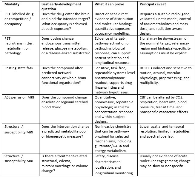 Table 1. Practical positioning of common neuroimaging modalities in an early CNS evidence package. PET nomenclature and ASL implementation should follow established quantitative standards.8,9
PET: molecular specificity with a narrow but powerful question
PET is the clearest imaging route to direct receptor, transporter, enzyme, or pathological-substrate measurements. A labelled investigational drug can establish biodistribution. More commonly, a selective radiotracer is administered before and after the test compound, and the reduction in tracer binding is modelled as occupancy. Linking occupancy to plasma exposure allows estimation of an EC50 or Emax relationship and supports dose selection. When the tracer is sensitive to endogenous neurotransmitter competition, PET can also quantify pathway activation - for example, dopamine release after a pharmacological challenge.
The strength of PET is not simply a colourful image. It is a quantitative model built around tracer kinetics, input or reference data, scan timing, and the biological meaning of binding potential.8 A poor tracer or poorly timed study can create false confidence. When no suitable radioligand exists, development teams should resist forcing PET into the programme and instead design a convergent package using CSF, peripheral target assays, MRS, fMRI, ASL, or electrophysiology.
MRI: repeatable functional pharmacology, with disciplined interpretation
MRI offers repeated measures without ionising radiation and can sample multiple aspects of central physiology in the same study. That makes it particularly useful for first-in-human dose escalation, crossover studies, temporal profiling, and pharmacological challenge models. But MRI-derived changes are typically one or more steps downstream from molecular binding. Their credibility therefore depends on a stronger experimental design: stable or well-characterised exposure, placebo and ideally positive controls, repeated scans spanning the PK profile, rigorous motion and physiological monitoring, and prespecified region or network hypotheses.3
The 2017 review led by Khalili-Mahani and colleagues surveyed approximately 85 acute pharmacological resting-state studies and found a diverse toolbox - functional connectivity, graph metrics, cerebral blood flow, and BOLD amplitude and spectral measures - but also major heterogeneity in acquisition, preprocessing, and biological-confound handling. The authors recommended multimodal datasets, sham-placebo or active controls, repeated psychometric and physiological measurements, and PK/PD modelling as the route from sensitivity to interpretable pharmacology.3
What controlled human pharmacology can add to CNS imaging
CHDR’s contribution to CNS imaging has focussed on embedding neuroimaging in controlled human pharmacology. Across studies with the Leiden University Medical Centre (LUMC) and other academic imaging partners, investigators have aligned dosing and scanning with PK, used placebo-controlled and crossover designs where feasible, collected functional and subjective readouts, and explicitly examined physiological confounding. This approach has been applied across several pharmacological imaging studies, including:
THC and PET: dynamic 18F-fallypride PET was used to measure THC-induced dopamine release, showing downstream pathway activation rather than CB1 receptor occupancy.7
Morphine, alcohol, and ASL: PK-controlled PCASL distinguished drug-specific cerebral blood flow patterns and showed the importance of accounting for respiratory and other physiological effects.4
Dopaminergic modulation and resting-state fMRI: haloperidol and levodopa produced network-specific and nonlinear connectivity effects, illustrating why fMRI endpoints should be hypothesis-led rather than treated as generic brain activation measures.5
Cross-compound fMRI analysis: resting-state connectivity changes across ethanol, morphine, THC, and ketamine were linked to a perception-related CNS phenotype, supporting the value of standardised imaging fingerprints.6
The common design lesson
Imaging becomes informative when it is embedded in the right experimental design. None of these studies relied on the scan in isolation. Each paired imaging with controlled perturbation, defined timing, quantitative analysis, and additional pharmacodynamic or behavioural information. The generalisable asset is not the modality alone, but the experimental medicine framework around it.
In practice, this means defining the decision, identifying the biological link that matters most, and building the evidence package around that link. The examples below show how this logic can be applied to three common CNS development scenarios.
Three common evidence packages
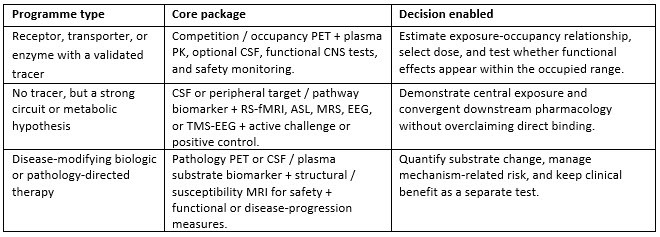 Table 2. Examples of convergent evidence packages. The exact context of use determines the required analytical validation, precision, and decision thresholds.1,2
Conclusion: use imaging to make the next decision
The strongest early CNS studies do not ask whether PET or MRI is "better." They ask what uncertainty blocks the next development decision and which measurement can remove it with the least ambiguity. PET is often the best tool for molecular distribution and occupancy. MRI is often the best tool for repeatable functional, perfusion, metabolic, and safety phenotyping. The most persuasive package links those readouts to exposure, challenge, physiology, and function.
CHDR’s body of work demonstrates this integration in practice: dynamic PET to quantify neurochemical pathway activation, PK-controlled ASL to separate drug-specific perfusion patterns, resting-state fMRI to reveal network-level and nonlinear pharmacology, and cross-compound analyses that connect imaging changes to subjective CNS effects. The common denominator is not the scanner. It is the experimental medicine framework around it.
For sponsors, the development standard should be simple: before advancing a CNS compound, be able to state to what extent the compound reached the brain, what target or pathway was engaged, how the relevant system changed, and why the selected dose is expected to matter. When neuroimaging is designed to answer those questions, it becomes more than a biomarker. It becomes a decision tool.
References
1. Morgan P, Van Der Graaf PH, Arrowsmith J, et al. Can the flow of medicines be improved? Fundamental pharmacokinetic and pharmacological principles toward improving Phase II survival. Drug Discovery Today. 2012;17(9-10):419-424. doi:10.1016/j.drudis.2011.12.020.
2. Vissers MFJM, Heuberger JAAC, Groeneveld GJ. Targeting for success: demonstrating proof-of-concept with mechanistic early phase clinical pharmacology studies for disease-modification in neurodegenerative disorders. International Journal of Molecular Sciences. 2021;22(4):1615. doi:10.3390/ijms22041615.
3. Khalili-Mahani N, Rombouts SARB, van Osch MJP, et al. Biomarkers, designs, and interpretations of resting-state fMRI in translational pharmacological research: a review of state-of-the-art, challenges, and opportunities for studying brain chemistry. Human Brain Mapping. 2017;38(4):2276-2325. doi:10.1002/hbm.23516.
4. Khalili-Mahani N, van Osch MJP, Baerends E, et al. Pseudocontinuous arterial spin labeling reveals dissociable effects of morphine and alcohol on regional cerebral blood flow. Journal of Cerebral Blood Flow & Metabolism. 2011;31(5):1321-1333. doi:10.1038/jcbfm.2010.234.
5. Cole DM, Beckmann CF, Oei NYL, Both S, van Gerven JMA, Rombouts SARB. Differential and distributed effects of dopamine neuromodulations on resting-state network connectivity. NeuroImage. 2013;78:59-67. doi:10.1016/j.neuroimage.2013.04.034.
6. Kleinloog D, Rombouts S, Zoethout R, et al. Subjective effects of ethanol, morphine, delta-9-tetrahydrocannabinol, and ketamine following a pharmacological challenge are related to functional brain connectivity. Brain Connectivity. 2015;5(10):641-648. doi:10.1089/brain.2014.0314.
7. Kuepper R, Ceccarini J, Lataster J, et al. Delta-9-tetrahydrocannabinol-induced dopamine release as a function of psychosis risk: 18F-fallypride positron emission tomography study. PLoS ONE. 2013;8(7):e70378. doi:10.1371/journal.pone.0070378.
8. Innis RB, Cunningham VJ, Delforge J, et al. Consensus nomenclature for in vivo imaging of reversibly binding radioligands. Journal of Cerebral Blood Flow & Metabolism. 2007;27(9):1533-1539. doi:10.1038/sj.jcbfm.9600493.
9. Alsop DC, Detre JA, Golay X, et al. Recommended implementation of arterial spin-labeled perfusion MRI for clinical applications: a consensus of the ISMRM Perfusion Study Group and the European Consortium for ASL in Dementia. Magnetic Resonance in Medicine. 2015;73(1):102-116. doi:10.1002/mrm.25197.