Supporting Phase 1 and first-in-human drug development
Targeted modulation of immune pathways has become the cornerstone of modern immunological drug development. Therapeutic strategies increasingly focus on cytokines, cytokine receptors, and intracellular signalling kinases that regulate innate and adaptive immune responses. A recent analysis in Nature Reviews Drug Discovery highlights sustained growth in programs directed at interleukins (ILs), tumour necrosis factor (TNF) superfamily members, and Janus kinase (JAK)–related pathways, alongside emerging upstream regulators such as thymic stromal lymphopoietin (TSLP), OX40/OX40L and TNF-like ligand 1A (TL1A). These targets represent a big part of current marketed products and late-stage clinical pipelines.
For early clinical development, human challenge models that activate these pathways provide an efficient translational bridge between preclinical studies and patient trials. CHDR has been at the forefront of developing and characterising controlled human inflammatory challenge models, including intravenous and intradermal lipopolysaccharide (LPS), keyhole limpet hemocyanin (KLH), and imiquimod (IMQ). The in-house availability of matched transcriptomic and proteomic datasets from these models represents a distinctive multi-omics capability that supports translational questions in early immunological drug development. Here, we evaluate the expression of clinically relevant immune targets within these challenge models and discuss their added value in early-phase clinical development.
Immunology drug target landscape: IL, TNF, JAK, and emerging pathways
Analysis of the current immunology drug landscape shows that the most prominent therapeutic targets cluster around a limited number of immune axes. These include cytokines and receptors from the IL-1, IL-2, IL-4/13, IL-5, IL-6, IL-12/23 and IL-17 families; TNF superfamily members such as CD40/CD40L, OX40/OX40L and TL1A; and intracellular signalling nodes including JAK1, JAK2, JAK3, TYK2 and IRAK4 (Figure 1). Together, these targets span early innate immune activation, T-cell and B-cell responses, and downstream amplification of inflammatory cascades. The breadth of this landscape emphasises the need for translational models that capture multiple immune mechanisms in humans.
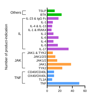 Figure 1. Immunology drug target landscape. Total number of drugs and development-stage agents across the 20 most prominent therapeutic targets in autoimmune and inflammatory diseases in 2024, grouped by pathway: TNF superfamily (TNF, TL1A, OX40/OX40L, CD40/CD40L), JAK kinases (TYK2, JAK1, JAK1/2, JAK1/2/3, JAK1 & TYK2), interleukins (IL-17, IL-5, IL-4, IL-6, IL-2, IL-1/IRAK4, IL-4/IL-13, IL-1, IL-23 & IgG Fc), and other kinase targets (BTK, TSLP). Each bar represents the number of product-indication pairs in 2024, counting multi-indication drugs once per indication. Pipeline counts include Phase II, Phase III, and regulatory filings not yet approved; marketed products are included and biosimilars and generics are excluded. Adapted from Fauconnier et al., Nature Reviews Drug Discovery (2025), based on Evaluate Pharma data.
Gene expression of immune targets in CHDR challenge models
Transcriptomic analyses across CHDR human inflammatory challenge models demonstrate altered expression of the majority of these key therapeutic targets (Figure 2). The models differ in their immunological profile, time course, and route of administration, and together provide complementary coverage of the therapeutic target landscape.
In the LPS challenge model, strong activation of innate immune pathways is observed, with prominent expression of IL-1, IL-6, TNF-related genes and downstream signalling kinases, including JAK family components. Uniquely, CHDR also profiled bone marrow in this model, capturing target expression at the primary site of innate immune cell production. The profile of the LPS challenge model closely aligns with targets pursued for modulation of acute inflammatory and cytokine-driven responses. The translational utility of the CHDR LPS challenge has been demonstrated in a proof-of-pharmacology study of an oral p38 MAPK inhibitor for the prevention of cancer immunotherapy-induced cytokine release syndrome. In a randomised, double-blind, placebo-controlled Phase 1 study conducted at CHDR, healthy participants received the investigational compound (30, 70, or 150 mg twice daily for seven days) and were challenged with both intradermal (ID) and intravenous (IV) LPS. Following ID LPS challenge, administration of the investigational compound markedly reduced local immune cell recruitment in suction blister fluid, with statistically significant suppression of neutrophils, classical monocytes, and CD3+ T cells. At the highest dose, treatment further significantly decreased local LPS-driven IL-1β and TNF levels in blister fluid compared to placebo. Following IV LPS challenge, p38 MAPK phosphorylation in CD14+ monocytes was significantly and dose-dependently decreased. Together, these results demonstrate that the CHDR LPS challenge model can quantifiably detect pharmacodynamic inhibition of inflammatory pathways in vivo, supporting its use in early-phase drug development (Figure 3A).
The KLH challenge model reflects antigen-driven adaptive immunity, and shows upregulation of targets associated with T-cell and B-cell activation, including CD40/CD40L and OX40/OX40L costimulatory pathways, IL-2 family cytokines (IL-2, IL-4, IL-21), JAK family kinases (JAK1, JAK2, TYK2), and BTK – a key kinase in B-cell receptor signalling. IL-23A is also upregulated, reflecting Th17 pathway engagement. This makes KLH a relevant model for evaluating therapies targeting adaptive immune mechanisms, including those in development for autoimmune indications.
The KLH model has been applied successfully in a first-in-human study of an anti-OX40L monoclonal antibody conducted at CHDR. In this Phase 1, randomised, placebo-controlled trial in 64 healthy male participants, intramuscular KLH immunisation followed by an intradermal KLH skin rechallenge was used to evaluate pharmacodynamic effects across single and multiple ascending doses. Administration of the investigational antibody suppressed the KLH-driven adaptive immune response in a dose-dependent manner, reducing skin erythema and cutaneous blood perfusion at the intradermal challenge site and systemic anti-KLH IgG antibody titers. This was the first study to establish the proof-of-pharmacology for OX40L blockade in humans (Figure 3B).
The IMQ challenge model is widely used as a translational model for psoriasis-like skin inflammation. It demonstrates pronounced upregulation of the TNF superfamily members (TNFSF13B, TNFSF15, TNF, TNFSF4), IL-family cytokines (IL-6, IL-10, IL-12B, IL-20, IL-36G), innate signalling kinases (IRAK4, RIPK1, RIPK2, MAP3K8, TYK2, JAK3), and T-cell activation mediators including ITK. IL-23A upregulation is also present, consistent with engagement of the IL-17/IL-23 axis which is relevant for psoriatic pathology. These expression patterns mirror key pathways targeted by current and emerging therapies in dermatology and other IL-17- and IL-23-driven autoimmune indications.
Protein-level confirmation across challenge models
To complement the transcriptomic findings, CHDR is able to assess the protein-level data using the Olink assay platform. This was performed for LPS challenge models (Figure 2). Matched proteomic datasets allow direct comparison of gene expression with circulating and compartment-specific protein levels, which strengthens the translational interpretation of the findings.
Proteomic data can also be derived from cerebrospinal fluid (CSF), which can provide a rare window into central nervous system compartment responses following systemic LPS challenge. The availability of matched RNA sequencing and Olink datasets within the same study designs represents a distinctive in-house multi-omics capability. This enables compound-specific questions about target expression, target engagement, and downstream pathway modulation to be addressed with a level of biological depth that is not routinely achievable in standard early-phase trial designs.
Translational value in de-risking immunology drug programs
Across CHDR challenge models, there is substantial overlap between expressed genes and the top immunology drug targets identified in current clinical pipelines. This concordance indicates that CHDR’s challenge models capture core immune mechanisms of high therapeutic relevance.
These datasets are available in-house and can be used directly to answer compound-specific questions. Transcriptomic and biomarker analyses can test whether a drug engages its target, modulates the expected pathway, or alters the inflammatory response. CHDR works with development teams to apply these models to the questions that matter for their programme.
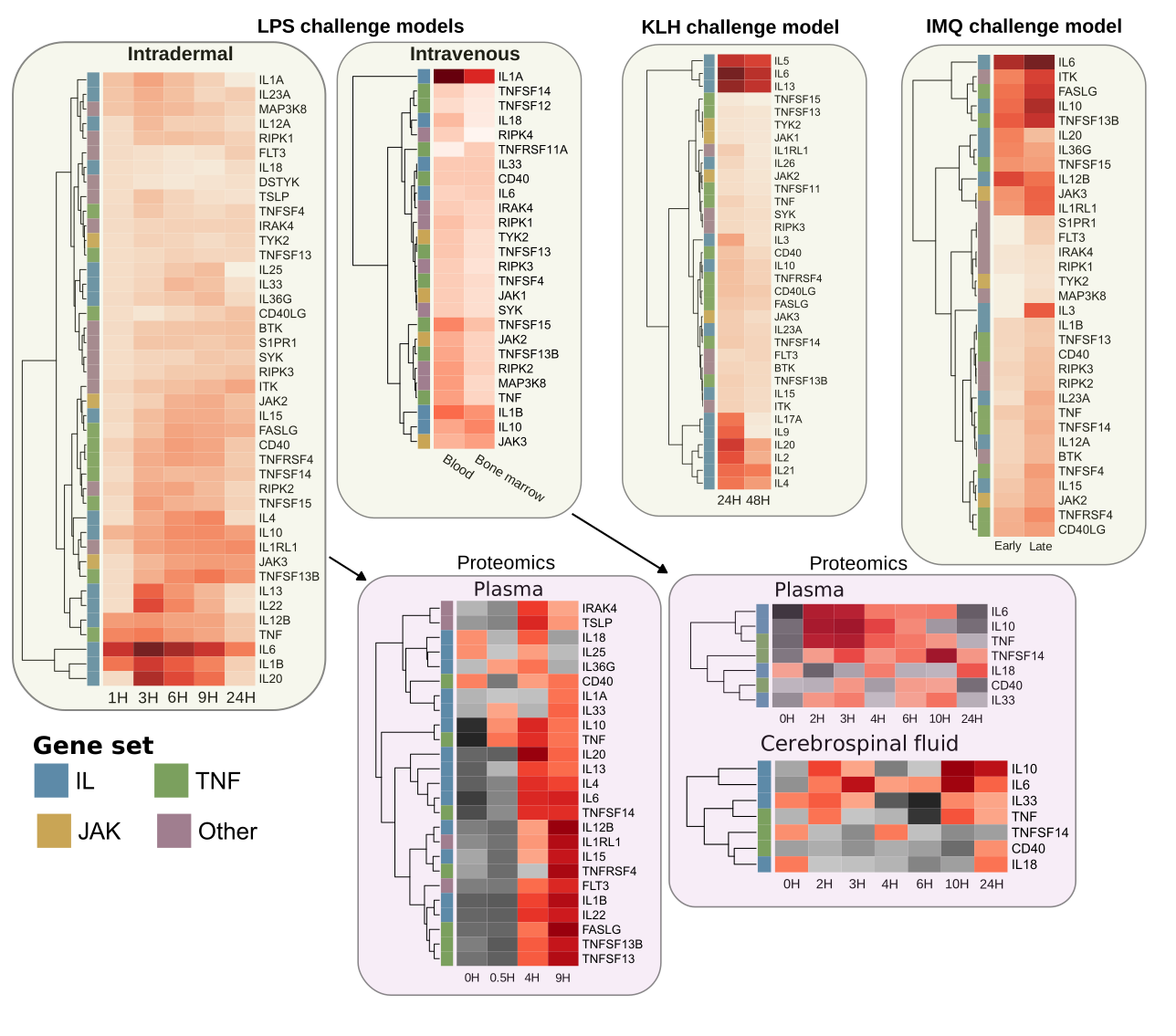 Figure 2. Upregulated immune targets across CHDR human challenge models. Heatmap panels showing transcriptomic (RNA sequencing, upper panels) and proteomic (Olink, lower panels) expression of key immunology drug target genes across CHDR human inflammatory challenge models. Genes are colour-coded by target family (IL, JAK, TNF, Other). Figures are schematic representations based on real data; absolute fold-change values and statistical thresholds are not shown.
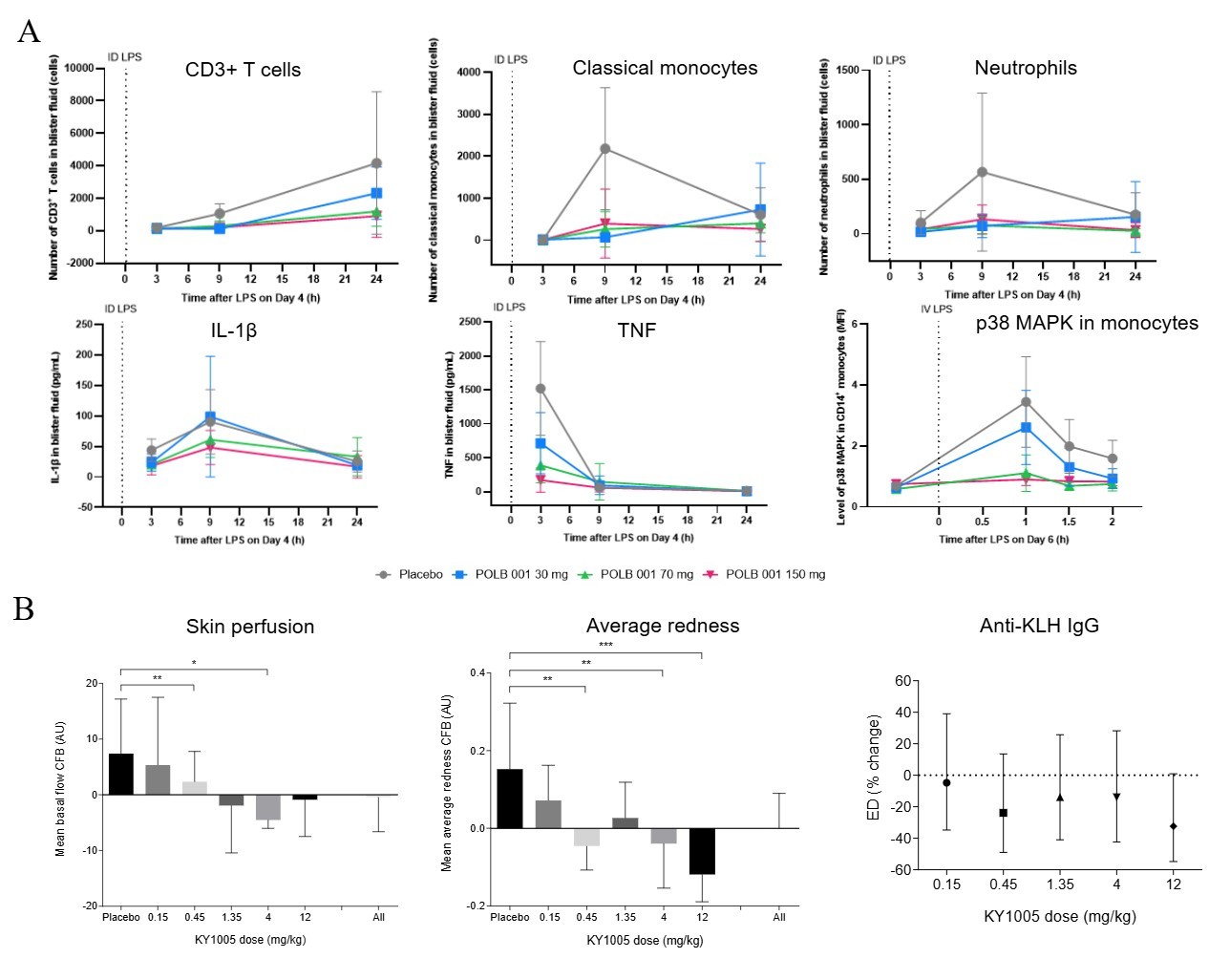 Figure 3. Pharmacodynamic suppression of inflammatory responses in CHDR human challenge models. (A) Effect of a p38 MAPK inhibitor on local and systemic inflammatory endpoints following LPS challenge in healthy participants. Upper panels: immune cell counts (CD3+ T cells, classical monocytes, neutrophils) in suction blister fluid following intradermal LPS challenge on Day 4, collected at 3, 9, and 24 h. Lower panels: cytokine levels (IL-1β, TNF) in blister fluid and p38 MAPK phosphorylation in CD14+ monocytes following intravenous LPS challenge on Day 6. Data are means ± SD. Adapted from de Bruin et al., Frontiers in Immunology (2025). (B) Effect of an anti-OX40L mAb on cutaneous skin perfusion, average redness, and anti-KLH IgG antibody titers following intradermal KLH skin rechallenge and intramuscular KLH immunisation, respectively. Data are shown as estimated differences (ED) from placebo ± 95% CI by dose group. p < 0.05, *p < 0.01, ***p < 0.001. Adapted from Saghari et al., Clinical Pharmacology & Therapeutics (2022).