Summary
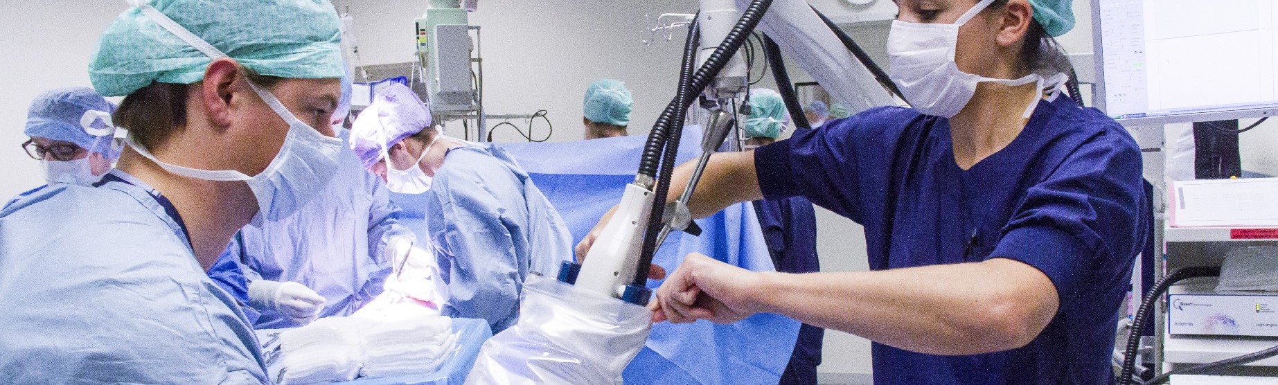
Image-guided surgery is a novel multidisciplinary technique that improves tumour diagnostics and increases the success of surgical cancer treatment. This technique can be used in classic ‘open’ surgeries, minimally invasive surgeries, and diagnostic procedures. A fluorescent label is attached to a molecule (e.g. a monoclonal antibody) that binds specifically and selectively to tumour cells, making the primary tumour and metastases visible to the surgeon. The fluorescent label itself is not pharmacologically active; however, it can be extremely useful for optimising the dosage and timing of pharmacological compounds.
The image-guided surgery team
Read more about the work of the image-guided surgery team on the LUMC website.
Practical answers to important research questions
-
What is the optimal dose for administering the labelled molecule in order to accurately visualise the tumour against background (i.e. healthy) tissue?
Using pharmacological measurements, CHDR can establish the optimal conditions for visualising tumour cells, metastases, and positive lymph nodes, thereby minimising both false-negative signals (which would result in incomplete removal of tumour cells) and false-positive signals (which would result in removal of healthy tissue).
-
What is the optimal timing for administering and tracking the fluorescent label?
After administration, the label must reach the tumour. Initially, healthy tissue may also absorb the label, temporarily becoming fluorescent. Depending on a variety of conditions, however, the label will be cleared from healthy tissue but will remain in the tumour cells, providing a window of time during which only tumour cells are fluorescent. CHDR uses sophisticated modelling techniques to determine the ideal timing between administering the label and performing the medical procedure.